Figure 10.
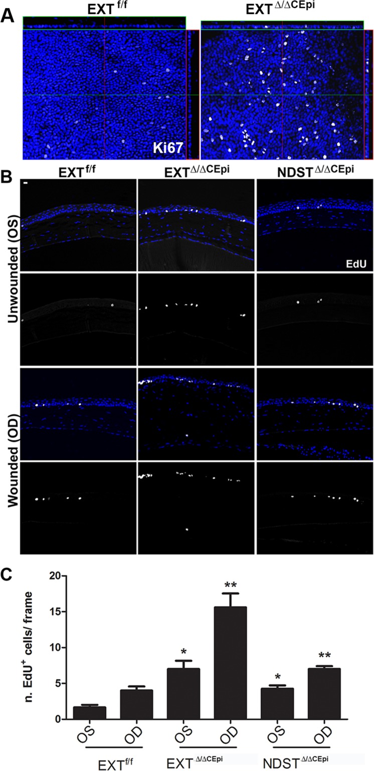
EXTΔ/ΔCEpi mice present increased cell proliferation. (A) Ki67 staining was performed on whole-mount corneas of P55 EXTf/f and EXTΔ/ΔCEpi mice induced at P21. (B) Debridement wounds were performed on P55 EXTf/f, EXTΔ/ΔCEpi, and NDSTΔ/ΔCEpi mice induced at P21 and mice submitted to EdU labeling 7 days after wounding for 4 hours. EXTΔ/ΔCEpi mice presented an increase in EdU incorporation in both the wounded and unwounded corneas. Corneas were processed for paraffin sectioning and analyzed by Click-IT Assay using Alexa 647. EdU labeling of sections 7 days after wounding (OD) EXTf/f, EXTΔ/ΔCEpi, and NDSTΔ/ΔCEpi mice compared to unwounded contralateral corneas (ES). (C) Number of EdU-positive cells in wounded and unwounded corneas of EXTf/f, EXTΔ/ΔCEpi, and NDSTΔ/ΔCEpi mice. Scale bar: 20 μm.

