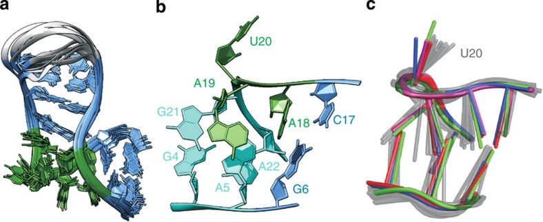
Figure 3. ssNMR structure of the Pf Box C/D RNA.
(a) Overlay of the 10 lowest energy structures of the Pf Box C/D RNA in complex with L7Ae from ssNMR data. Terminal nucleotides 1 and 25–26 are not shown. Colour code as in Fig. 1a. (b) k-turn of the Pf Box C/D RNA, showing the characteristic geometry. Internal loop, green; NC stem, cyan, C stem, light blue. (c) Comparison of the k-turn geometry of the Pf Box C/D RNA obtained by ssNMR (10 lowest energy structures, gray) with that of the crystallographic structure of the Af Box C/D RNA (PDB code 1RLG)7, red; Pf Box C/D RNA (PDB code 3NMU)8, blue; Af Box C/D RNA (PDB code 4BW0)29, green; Ss Box C/D RNA (PDB code 3PLA)30, magenta.

