Figure 3.

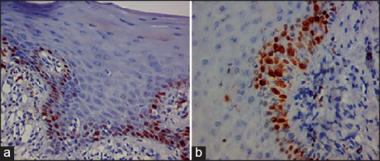
Immonohistochemical view (×20) (a) P53 and (b) Ki-67. Showing nuclear brown staining of tumor markers in basal and parabasal cells of oral epithelial dysplasia.

Immonohistochemical view (×20) (a) P53 and (b) Ki-67. Showing nuclear brown staining of tumor markers in basal and parabasal cells of oral epithelial dysplasia.