Fig. 2.
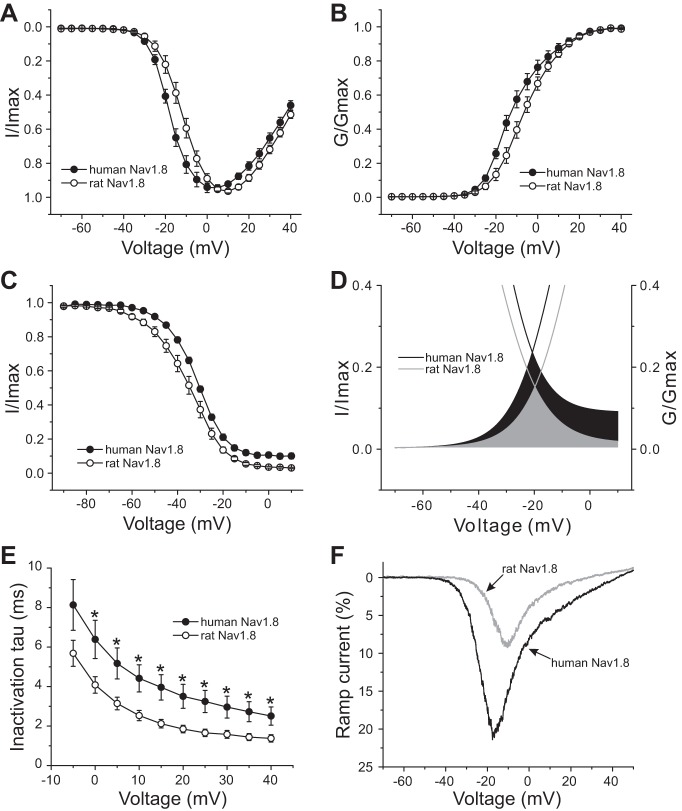
Voltage-clamp analysis of human and rat Nav1.8 channels in transfected Nav1.8-knockout mouse DRG neurons. A: normalized current-voltage (I–V) curves for human and rat Nav1.8 channels. B: comparison of voltage-dependent activation between human and rat Nav1.8 channels. Activation of human Nav1.8 channels is hyperpolarized by ∼5 mV compared with rat Nav1.8 channels. G/Gmax, normalized sodium conductance. C: fast inactivation of human Nav1.8 is depolarized by 3.7 mV compared with rat Nav1.8. D: magnified view of overlap between activation and fast inactivation for rat and human Nav1.8. The overlap of Nav1.8 channels for rat is shown in gray, whereas the black area shows the difference of the overlap between human and rat Nav1.8. E: kinetics of open-state inactivation measured as a function of voltage for rat (n = 17) and human (n = 17) Nav1.8 channel. Time constants (τ) were obtained by fitting currents elicited as described in Fig. 1, A and B, with a single-exponential function. *P < 0.05. F: representative ramp currents elicited with 600-ms ramp depolarization from −70 mV to 50 mV for human or rat Nav1.8 channel recorded from transfected DRG neurons. Human Nav1.8 produces a ramp current that is almost double that produced by rat Nav1.8 channels.

