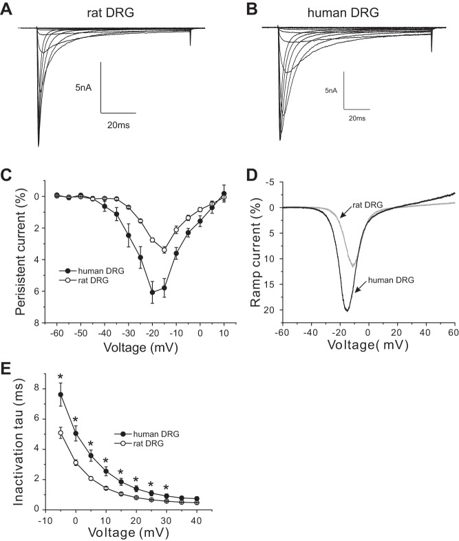
Fig. 7.
Human DRG neurons demonstrate larger Nav1.8 persistent currents. A and B: representative Nav1.8 current family traces recorded from a rat DRG neuron (A) or a human DRG (B). Cells were held at −60 mV and stepped to a range of potentials (−60 to +40 mV in 5-mV increments) for 100 ms. C: comparison of the normalized amplitude of Nav1.8 persistent currents between rat DRG neurons and human Nav1.8 DRG neurons. D: representative ramp currents, elicited with 600-ms ramp depolarization from −60 mV to +60 mV, for Nav1.8 channel in a human and a rat DRG neuron. Nav1.8 channels in human DRG neurons produce a ramp current that is almost double that produced by Nav1.8 channels in rat DRG neurons. E: kinetics of open-state inactivation measured as a function of voltage for Nav1.8 channel in rat DRG neurons (n = 19) and in human DRG neurons (n = 9). Time constants were obtained by fitting currents elicited as described in A and B with a single-exponential function. *P < 0.05.

