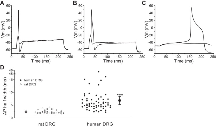
Fig. 8.
Human DRG neurons generate broader action potentials. A and B: representative action potential traces recorded from a rat (A) or a human (B) DRG neuron. Action potentials were elicited by 200-ms step depolarizing current injections from resting membrane potential. C: action potential traces recorded from a human DRG neuron with prolonged duration. D: comparison of the half-width of action potential between rat and human DRG neurons. Two larger symbols indicate means ± SE. ***P < 0.001.

