
FIG. 8. a, b.
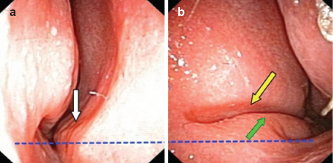
An anterior, basal crest can be seen (white arrow). In most cases, it touches the mucosa of the inferior turbinate. This photograph was taken after the decongestion and superficial local anesthesia; thus, much closer contact between the crest and inferior turbinate can be presumed and imagined (a). The typical septal groove is located on the opposite septal side (yellow arrow); intermaxillary bone wing contours are clearly presented (green arrow) (b). Blue dotted line shows that the left nasal floor stays at the higher level than the right one: this is very typical for type 6.