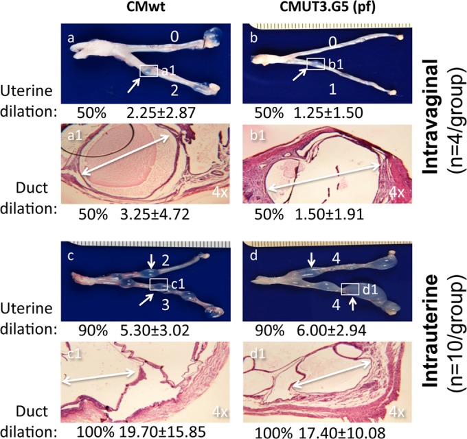
FIG 6.

Effect of plasmid depletion and infection routes on C. muridarum induction of glandular duct dilation. Four groups of C57BL/6J mice, with 4 mice in each of the first two groups (a, a1, b, and b1) and 10 mice each in the third and fourth groups (c, c1, d, and d1), were infected with wild-type C. muridarum (CMwt) (a, a1, c, and c1) or the plasmid-free C. muridarum clone CMUT3.G5 (pf) (b, b1, d, and d1) either intravaginally (a, a1, b, and b1) or intrauterinally (c, c1, d, and d1). Sixty days after infection, mice were sacrificed to observe upper genital tract pathology, as described in Materials and Methods. For uterine horn dilation under the naked eye, the white arrows indicate a representative dilated area from each horn, and the dilation severity score for each uterine horn is shown by a white number. To examine glandular duct dilation, the uterine horn dilation lesion areas (boxed and labeled a1 to d1) were subjected to microscopic observation under a 4× objective lens. One representative dilated glandular duct is marked with a white double-headed arrow in each representative H&E image. Both the incidence rates and severity scores of either uterine horn or glandular duct dilation from each group are shown under the corresponding images. Note that 50% or more of the mice developed significant uterine horn and glandular duct dilation regardless of the plasmid deficiency (compare panels a and a1 with b and b1 or c and c1 with d and d1). Intrauterine infection did increase both the incidence rates and severity scores of the dilation (compare panels a and a1 with c and c1 or b and b1 with d and d1).
