FIG 7.

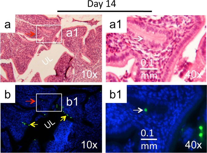
Immunohistochemical detection of plasmid-free C. muridarum in glandular duct epithelial cells. C57BL/6 mice intravaginally infected with the plasmid-free C. muridarum were sacrificed on day 14 after infection for immunohistochemical detection of C. muridarum in the genital tract tissues. (a) The H&E staining images taken under a 10× objective lens revealed the overall structure of the uterine horn, with the horn lumen (UL) marked. (b) The same section was immunofluorescence labeled with an antibody against C. muridarum organisms (green) and the Hoechst DNA dye (blue) prior to H&E staining. C. muridarum organisms were detected along both the uterine horn lumenal epithelial cells (yellow arrows) and glandular duct epithelial cells (green inclusions indicated by red arrows). (a1 and b1) A representative glandular duct positive for C. muridarum labeling (boxes in panels a and b) was selected for further observation under a 40× objective lens. The location of the inclusion marked with a white arrow in the immunofluorescence-labeled slide (b1) is indicated in the H&E slide (a1).
