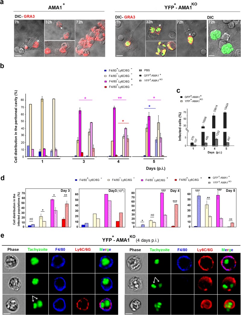
FIG 3.
AMA1KO and AMA1+ tachyzoites colonize resident and inflowing immune cells. (a) Cells from the peritoneal fluids of mice inoculated with 105 DiCre YFP− AMA1+ (left panel) or 105 or 106 DiCre YFP+ AMA1KO (right panel). The GRA3 protein was detected in growing parasites and in their surrounding PV/PVM (red; the white arrowheads point to the vacuoles, while white arrows point to the large vacuoles frequently seen with the 106 inoculum). (b to e) AMNIS analysis of cells from peritoneal exudates of mice inoculated with 105 GFP+ AMA1+ or YFP+ AMA1KO and labeled for F4/80 and Ly6C/6G. (b) Relative distributions of cell subpopulations while mutant and wild-type tachyzoites are fluorescent. (c) Percentages of infected cells, with the numbers of cells analyzed indicated on top of each column. (d) Relative distributions between the infected cell subpopulations. (e) Images for each type of peritoneal cell infected by AMA1KO parasites at day 4 p.i. Bars, 10 μM.

