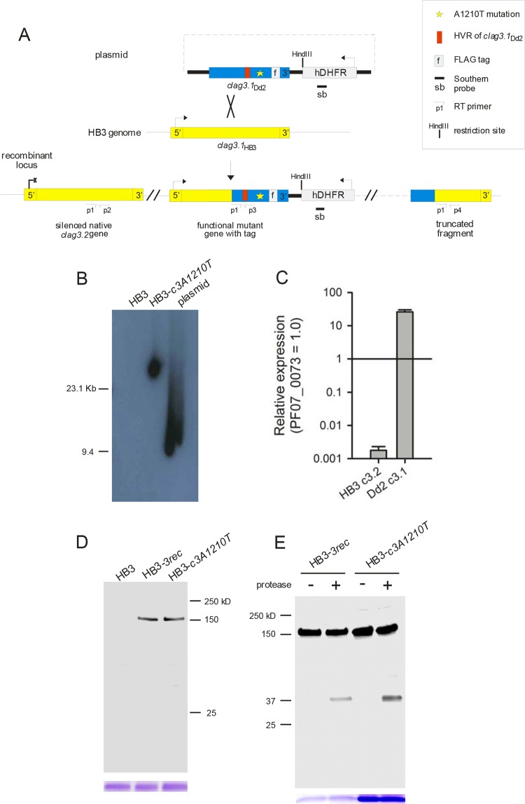
FIG 2.
Allelic-exchange transfection and expression of CLAG3 carrying the A1210T mutation. (A) Schematic showing the integration plasmid (top) and the result of allelic-exchange transfection to replace the wild-type HB3 clag3.1 gene with Dd2 clag3.1 carrying a nonsynonymous mutation encoding threonine at residue 1210. The positions of the primers (see Materials and Methods) and the probe used for Southern blotting are shown. (B) Southern blot assay with genomic DNA from the clones indicated or the integration plasmid after HindIII digestion. Plasmid size, 9.4 kb. While the hdhfr-specific probe does not recognize DNA from HB3, a single integration site is apparent in HB3-c3A1210T. (C) Mean ± the standard error of the mean transcript abundance of the genes indicated in HB3-c3A1210T from RT-PCR experiments with normalization to the PF07_0073 control. (D) Immunoblot assay of whole-cell lysates of cells infected with the parasites indicated probed with anti-FLAG antibody. A single band of the expected size (∼150 kDa) is apparent in HB3-c3A1210T and HB3-3rec. Coomassie blue staining of hemoglobin (used as a loading control) is shown at the bottom. (E) Immunoblot assay of the membrane fraction harvested from cells with or without extracellular pronase E treatment. Anti-FLAG antibody recognizes a C-terminal ∼37-kDa cleavage product in both transfectants after pronase E treatment. Loading control, Coomassie blue staining of hemoglobin.

