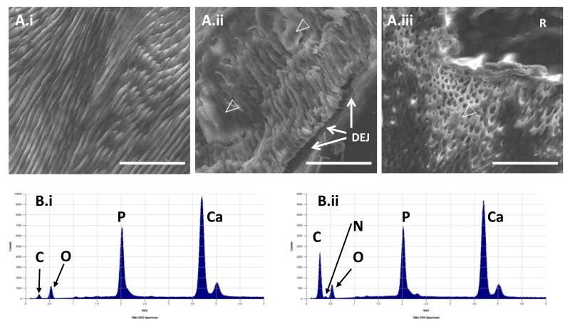
Figure 2. Ultrastructural phenotyping of deciduous teeth.
A) SEM of exfoliated deciduous tooth cervical enamel. The appearances of control enamel (i) contrast with the poorly formed enamel rods that are partially obscured by amorphous material (open arrow heads) in affected incisor (ii) and molar (iii) teeth. R is embedding resin and not dental tissue; bar 50μm. B) EDX spectra. In control enamel (i), the C:O ratio is low compared to that in affected enamel (ii). A small N peak was observed (between the C & O peaks) in affected, but not control teeth. Similar peaks are observed in affected and control teeth for Ca and P.

