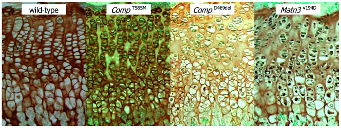
Figure 1.
Organization of the growth plate in mutant mice is disrupted by 3 weeks of age and shows marked hypocellularity along with the retention and/or mislocalisation of cartilage structural proteins. Representative immunohistochemistry (IHC) of the growth plates from 3 week-old wild-type and mutant mice showing disruption to chondrocyte columns in mice homozygous for the Comp p.Thr585Met (CompT585M), Comp p.D469del (CompD469del) and Matn3 Val194Asp (Matn3V194D) mutations. IHC using COMP (wild-type, CompT585M and CompD469del) and matrilin-3 (Matn3V194D) antibodies revealed less staining in the extracellular matrix (ECM) between the proliferating columns in the growth plates of mice carrying all three mutations. Furthermore, there was intracellular staining for mutant COMP and matrilin-3 in chondrocytes from the CompD469del and Matn3V194D mice, respectively.

