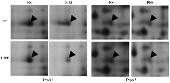
Figure 3.
Two-dimensional gel electrophoresis patterns of protein expression in rat brains for two selected proteins. FC, prefrontal cortex; Hipp, hippocampus; NS, non-prenatal stressed offspring; PNS, prenatal stressed offspring. Arrowheads are protein spots, showing different changes among the groups.

