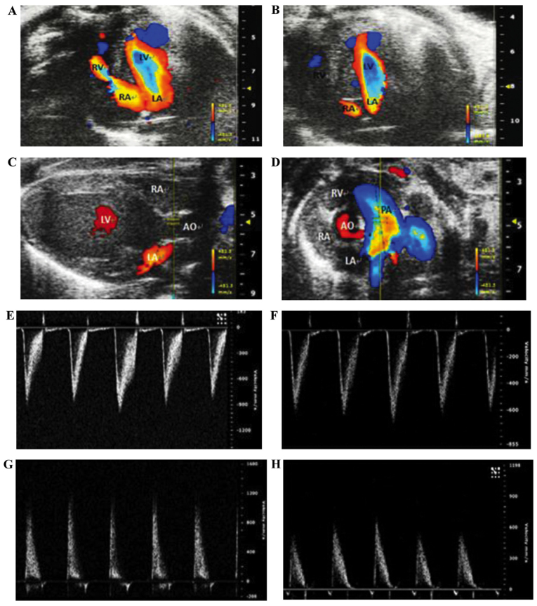
Figure 5.
Echocardiography showing an atrial septal defect (ASD) in the heterozygous transgenic mice: (A–D) color Doppler echocardiographic images; and (E–F) pulsed Doppler echocardiographic images. (A) An apical four chamber view of the heterozygous mouse heart, displaying the blood circulation between the atria. (B) An apical four chamber view of the wild-type mouse heart, with no obvious abnormality. (C and D) Images of pulmonary valve peak velocity of the heterozygous transgenic mouse and its wild-type littermate, respectively. (E and F) The pulsed Doppler echocardiographic images across the pulmonary valves of the heterozygous transgenic mouse and its wild-type littermate, respectively. (G and H) The pulsed Doppler echocardiographic images across the aortic valves of the heterozygous transgenic mouse (G) and its wild-type littermate (H). RV, right ventricle; LV, left ventricle; RA, right atrium; LA, left atrium; AO, aorta.

