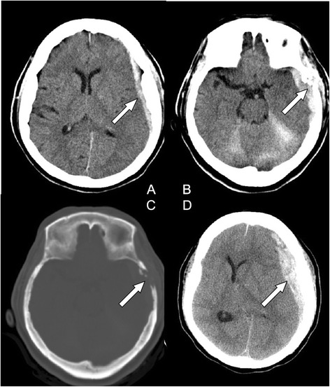
Fig. 1.

(a) Computed tomography (CT) of the head showing left temporal subdural hematoma (SDH) (arrow) with slight mass effect. (b) small left temporal bone erosive lesion (arrow). (c) A bony defect on the left temporal bone (bone window of CT) (arrow). (d) Enlargement of left SDH with significant mass effect (arrow)
