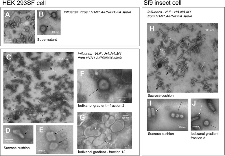
Figure 1.

NSTEM images at 40,000× magnification of H1N1 A/Puerto Rico/8/34 (A & B), H1N1 influenza-VLPs from HEK 293SF cell production (C-G) or Sf9 cell production (H-J). VLPs or influenza virus are pointed by black arrows, baculovirus and cell vesicles are pointed by white arrows. A & B - Supernatant of H1N1 A/Puerto Rico/8/1934 influenza virus produced in HEK 293SF suspension cells as in Petiot et al. [28]. Cell vesicles carrying influenza glycoproteins could be identified. C, D & E - Sucrose cushion purified samples of HEK 293SF cell production; large view (C) and zoom-in (D & E) images of NSTEM grid. F & G - Iodixanol purified samples of HEK 293SF cell production. Typical shapes of VLPs and vesicles identified in high density (fraction 12: 1.03 g/ml) and low density (fraction 2:) iodixanol fractions. H & I - Sucrose cushion purified samples of Sf9 cell production. J - Iodixanol purified samples fraction 3, which present the highest number of VLP particles. Baculovirus were co-purified with VLPs in all the iodixanol fractions, even if they were more concentrated in the high density fractions.
