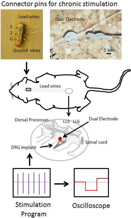
Figure 1.
Diagrammatic overview of the stimulation surgery. Dual electrodes were implanted into the DRG at L5 or L6. Lead wires ran from the electrodes up the back of the animal to an adaptor affixed to the skull. This head fixture allowed for repeated stimulation and included ground, electrode 1 (1) and electrode 2 (2) connections. Animals were subjected to 10 days of 400 μs pulses at 20 μAmps at 200 Hz for 1 hour/day and the stimulation paradigm monitored with an oscilloscope.

