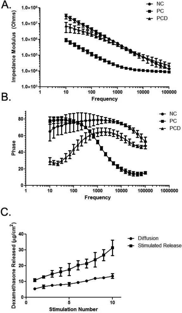
Figure 3.
In vitro impedance spectroscopy for NC, PC and PCD conditions. A: Bode plots. The lowest Z values were observed with the PC coating (solid squares) and incorporation of dexamethasone resulted in an impedance spectrum for the PCD coating (solid triangles) more similar to that observed with NC (solid circles). B: Phase plot indicating the different electrode-electrolyte interface behaviors for NC (solid circles), PC (solid squares) and PCD (solid triangles) coatings. Each plot includes data from n = 3 replicates, and error bars represent SD. C: Summed in vitro dexamethasone release. Over the ten stimulations applied over the course of the in vivo study, dexamethasone was released with electrical stimulation (solid squares). Electrical stimulation triggered significantly more (p < 0.0001) drug release than that observed with passive diffusion (solid circles).

