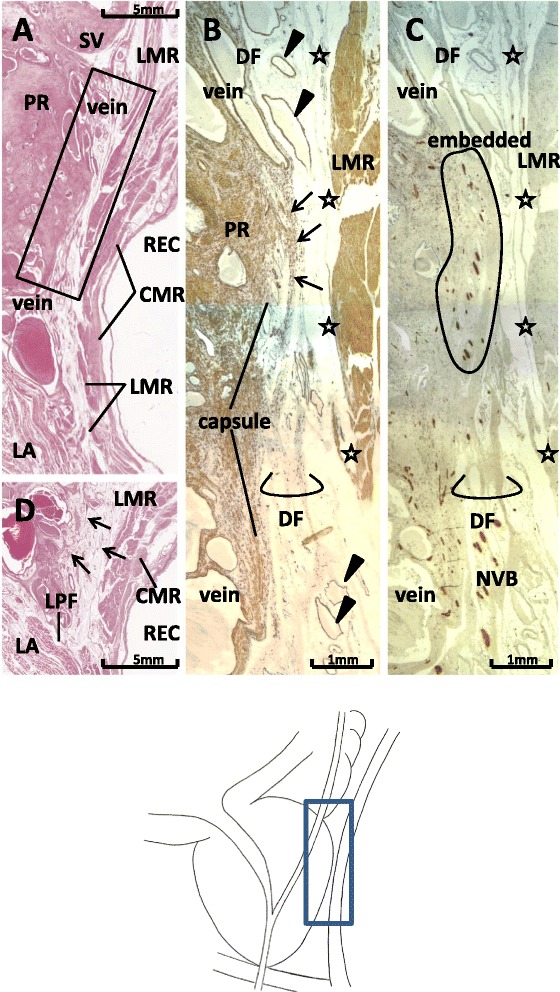
Figure 3.

Fascial complex between Denonvilliers’ fascia and the prostatic capsule in sagittal sections. Sections from a 75-year-old man are shown. Panel A (HE staining) displays topographical anatomy around Denonvilliers’ fascia (DF) at the 2- to 3-o’clock position of the rectum. The levator ani muscle (LA) approaches the rectum (REC). Panel B (Panel C), corresponding to a square in panel A, shows immunohistochemistry for smooth muscles (for all nerves). DF, containing smooth muscles, comprises 3–4 leaves (lower part of panel B), but the leaves are bundled to fuse with the prostatic capsule (capsule) in the upper part of the panel (arrows). At the fusion area (panel C), periprostatic nerves are embedded in the fascial complex between DF and the prostatic capsule (encircled). Stars indicate a candidate for the fascia propria of the rectum. The DAKO antibody that was applied for smooth muscles also strongly stains vascular endothelium (arrowheads). In panel D, 4 mm lateral to the area shown in panel A, fascial leaves (arrows) become thinner and fewer in number. CMR and LMR: circular and longitudinal muscle layers of the rectum; LP: lateral pelvic fascia; NVB: neurovascular bundle at the posterolateral corner of the prostate; PR: prostate; SV: seminal vesicle.
