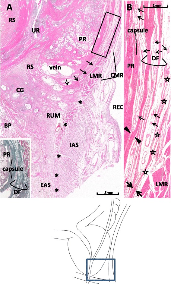
Figure 4.

Multiple-leaf configuration of Denonvilliers’ fascia in a midsagittal section from a 79-year-old man. Panel A displays topographical anatomy around Denonvilliers’ fascia (DF). The longitudinal muscle layer of the rectum (LMR) extends inferiorly to continue to the conjoint muscle coat or longitudinal anal muscle (asterisks). The leaves of DF (arrow) are dispersed in a venous plexus (veins) without ending at the rectourethralis muscle (RUM) in this specimen. The insert in the panel A (elastic–Masson staining; a section near panel A) shows rich content of elastic fibers (black color) in the prostatic capsule, DF, and fascia propria of the rectum. Panel B, corresponding to the rectangle in panel A, shows the multiple-leaf configuration of DF. Periprostatic nerves (thin arrows) are scattered between the fascial leaves. A thick leaf of the fascia is fused with the prostatic capsule (capsule) at the site indicated by arrowheads, while the other leaf merges with the fascia propria of the rectum (stars) at a site indicated by thick arrows. BP: bulbus penis; CG: Cowper’s gland, CMR: circular muscle layer of the rectum; EAS: external anal sphincter; IAS: internal anal sphincter; PR: prostate; REC: rectum; RS: rhabdosphincter area; UR: urethra.
