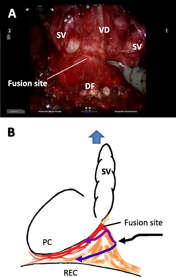
Figure 5.

Intraoperative findings (A) and schema (B) of Denonvilliers’ fascia. When the seminal vesicles and vas deferens are pulled ventrally, Denonvilliers’ fascia (DF) is observed as a membranous structure, and the fusion site of DF with the prostatic capsule is recognized near the seminal vesicle-prostate junction (Panel A). DF between the seminal vesicles and rectum should first be cut at the midline to avoid entering the prostatic capsule (black arrow). After this incision, a mesh-like structure is found behind the posterior aspect of the prostate. With a nerve-sparing procedure, the dissection plane should be as close to the prostate as possible (purple arrow). If there is advanced cancer at the border of the posterior aspect of the prostate, the dissection plane can remain adjusted to the rectal wall (blue arrow). PC: prostatic capsule; REC: rectum; SV: seminal vesicle; VD: vas deferens.
