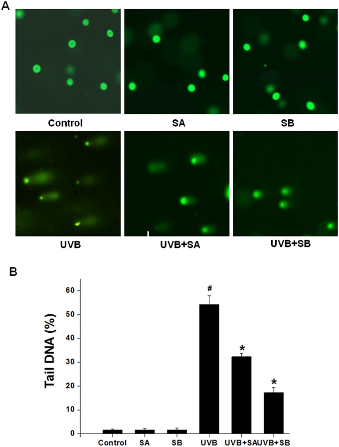
Fig 3. SA and SB reduced the DNA damage that was induced by UVB in HaCaT cells.
HaCaT cells were treated with 100 μM SA or SB for 24 h, followed by exposure to 30 mJ/cm2 of UVB. DNA damage was assessed using a comet assay at 24 h after UVB treatment. The percent of tail DNA was analyzed. (A) Representative epifluorescence microscopy images, 200× magnification. (B) Quantification of the fluorescence intensities of the images in panel A. #P < 0.05 was considered as a significant difference compared with the control group. *P < 0.05 was considered as a significant difference compared with the only UVB-irradiated group.

