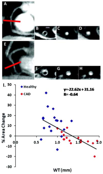
Figure 3.

Noninvasive assessment of early coronary atherosclerosis, coronary endothelial function, and their relationship with MRI. (Upper Panel) Typical anatomic and functional coronary images using MRI at rest and with isometric handgrip stress. In a healthy subject (A), a scout scan obtained along the right coronary artery (RCA) in a healthy adult subject is shown together with the location for cross-sectional imaging (red line). B, A view perpendicular to the RCA in a healthy adult subject is shown, illustrating a black blood vessel wall cross-section. C, A white blood view for endothelial function in a healthy subject is shown at rest (C) and during stress (D). In a patient with CAD (E–H), a scout scan obtained parallel to the RCA is shown (E) together with the location for cross-sectional imaging (red line). F, RCA black blood vessel wall cross-sectional image in the same patient with CAD. G, A view perpendicular to the RCA in the patient with CAD is shown at rest (G) and during stress (H). (Lower Panel). Relationship between early atherosclerosis and endothelial function is shown in the summary data (I). MRI measures of coronary wall thickness (mm) versus percent coronary cross-sectional area changes with isometric handgrip stress in healthy subjects and patients with coronary artery disease (combined) while individual data points are shown for healthy subjects (blue diamonds) and patients with coronary artery disease (red diamonds). Reproduced with permission from Hays et al (Hays et al. 2012)
