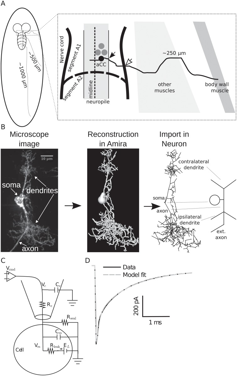
Fig 3. Morphological reconstruction of the 3rd instar aCC motoneuron.
(A) Schematic muscle projections of aCC motoneurons based on their location in the nerve cord segment. (B) Stack of microscope images was reconstructed using Amira software (Visage Imaging GmbH, Berlin, Germany) and then imported into the Neuron simulator [72]. The rightmost schema indicates the major morphological components in an idealized depiction. “ext. axon” indicates the missing extended axon from the reconstruction (not drawn to scale). (C) Equivalent circuit of the measured passive properties including an electrode model for voltage clamp. (D) Passive response to voltage-clamp step to −90 mV from a holding potential of −60 mV is simulated in Neuron with the fitted parameters (see Table 2).

