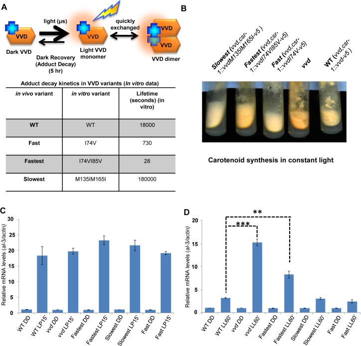
Fig 1. Altering VIVID (VVD) photocycle through site-directed mutagenesis reveals a photoadaptation mutant.
(A) Cartoon showing in vitro photochemistry of VVD light-activated dimer and the influence of specific mutations on the photo-adduct stability/life-time [20]. Blue crosses represent chromophores, and light activated chromophores are shown with orange halo. (B) WT, vvd null and three photocycle length variants were grown on solid minimal medium slants, exposed to constant light (40 μM m-2s-1) for 4–5 days and the mutants visually compared to the WT for carotenoid biosynthesis. (C) Strains (n = 3) exposed to a 15 minute light pulse (LP15’) were subjected to RT-PCR to determine al-3 gene expression levels as a measure of the integrity of the light response in the photocycle mutants. (D) Strains (n = 3) were exposed to bright white light for 60 minutes (LL60’) to study photoadaptation response using al-3 gene expression as a readout. The fastest photocycle mutant shows a partial loss of photoadaptation at the gene expression level when compared to the WT strain as seen by the higher levels of al-3 mRNA after 60 minutes of light exposure (LL60’). Asterisks indicate statistical significance as determined by an unpaired t test. **P<0.01, ***P<0.001.

