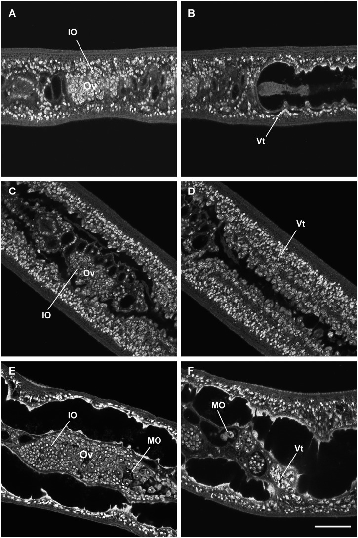
Fig 4. Confocal laser scanning microcopy images of female S. japonicum from unisexual infections cultured in vitro.
Females before cultivation (A, B), females cultured for 21 days in medium RPMI-1640 without RBCs (C, D) or with RBCs (E, F). Ov, ovary; IO, immature oocyte; MO, mature oocyte; Vt, vitelline cells. (A) Ovary containing numerous immature oocytes. (B) Undeveloped vitelline gland. (C) Ovary with immature oocytes. (D) Immature vitelline cells. (E) Enlarged ovary with mature oocytes. (F) Mature vitelline cells. Scale bar, 50 μm.

