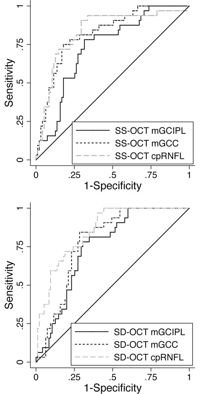
Fig 3. Receiver of operating characteristic curves showing the ability of mGCIPL, mGCC, and cpRNFL measured in swept source optical coherence tomography and spectral domain optical coherence tomography to distinguish healthy and glaucomatous eyes.

Areas under the receiver operating characteristic curve (AUC) of mGCIPL, mGCC, and cpRNFL were 0.73, 0.82 and 0.83 by swept source optical coherence tomography (SS-OCT), respectively (top), and 0.75, 0.78, and 0.85 by spectral domain optical coherence tomography (SD-OCT), respectively (bottom).
