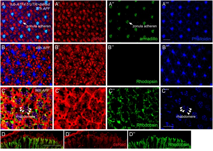
Fig 5. In vivo ATF4 reporter is activated in the photoreceptor cells.
(A) At 37% pupal development (37h APF), retina were stained with anti-dsRed (red), anti-armadillo (arrowhead, marker for zonula adherens, green), and anti-actin-phalloidin (blue), respectively. DsRed is expressed at this stage. (B) At 48% of pupal development (48h APF), ATF4 reporter was still active, but, rhodopsin was not detected at this stage. (C) At 96% pupal development retina (96h APF), ATF4 reporter activity was detected at the photoreceptor cells. (D) The expression of dsRed in the adult fly retina, but not in the photoreceptor. Red indicates ATF4 reporter and green is rhodopsin staining. The scale bar in (A′′′) represents 5 μm for (A-C) and that in D represents 50 μm.

