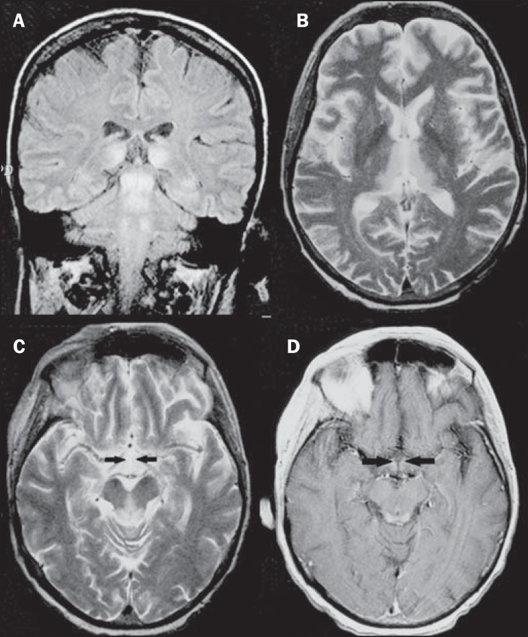
Figure 1.
Wernicke syndrome. Female, 47-year-old patient. Coronal FLAIR (A) and axial T2-weighted (B,C) images show hypersignal foci in the periacqueductal gray substance, thalami in paramedian region, mammillary bodies (arrows), tectum and tegmentum of the mesencephalon. Contrast-enhanced T1-weighted image (D) demonstrates enhancement of mammillary bodies (arrows) and tectum of the mesencephalon.

