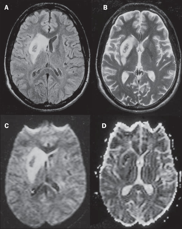
Figure 6.
Ischemic stroke/arteritis with infarction caused by use of cocaine. Male, 21-year-old patient. Lesions are present in the putamen, caudate nuclei head and anterior leg of the internal capsule at right. Presence of area of hypersignal on T2-weighted FLAIR image, with a focus of putaminal hyposignal intensity (hemorrhage) (A,B). Diffusion-weighted image demonstrates hypersignal (C) and at the ADC map (D) there is hyposignal, except on the putaminal area of hemorrhagic transformation, with hyposignal on diffusion-weighted image and on the ADC map.

