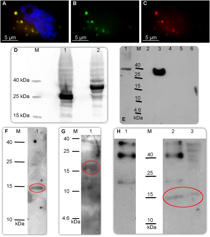
Fig 2. Molecular studies on CRNDEP.
A) Simultaneous overexpression of the 6xHis-CRNDEP-EGFP and DsRed Monomer-6xHis-CRNDEP fusion proteins in HeLa cells, visualized under a fluorescence microscope. The former protein glows green and the latter glows red in these conditions. Yellow glow is caused by a co-localization of these two fusion proteins. Nuclei were stained blue with DAPI. The same shot with only the green (B) or the red (C) channel shown. D) Western blot-based verification of the size of the 6xHis-CRNDEP-EGFP fusion protein. M—Spectra Multicolor Low Range Protein Ladder (Thermo-Fisher Scientific), 1—the EGFP reporter protein (26.9 kDa), 2—6xHis-CRNDEP-EGFP (39.2 kDa). E) Western blot-based verification of the specificity of our custom-made polyclonal anti-CRNDEP antibody. M—Spectra Multicolor Low Range Protein Ladder; 1—DsRed Monomer-6xHis-CRNDEP (340 aas, 38.5 kDa); 2—purified 14 kDa protein containing the 6xHis tag, 1.4 μg (a negative control of the antibody's specificity, non-commercial); 3—6xHis-CRNDEP-EGFP (346 aas, 39.2 kDa); 4—empty; 5—EGFP (239 aas, 26.9 kDa, a negative control); 6—DsRed Monomer (232 aas, 26.2 kDa, a negative control). A loading control (the PVDF membrane used in this experiment, stained with Ponceau S) is shown in S13 Fig. F–G) Detection of the overexpressed 2xFLAG-CRNDEP protein (~14 kDa) in a total protein lysate from 0.25 million HeLa cells with either the anti-FLAG (F) or anti-CRNDEP (G) antibody. H) Immunoprecipitation of 2xFLAG-CRNDEP using the anti-CRNDEP antibody (2) and control IgG (1) (both from a rabbit). A total protein lysate before immunoprecipitation was loaded for comparison (3). After precipitation, the 2xFLAG-CRNDEP protein was detected on the PVDF membrane using the anti-FLAG antibody. The correct bands in Fig F–H are encircled.

