Fig. 1.

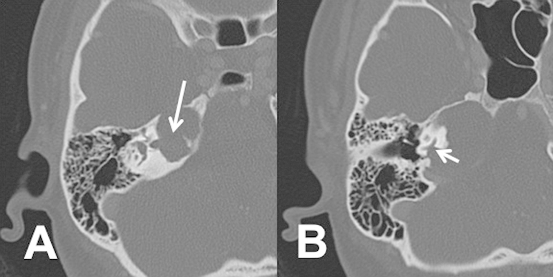
Axial computed tomography scan showing (A) internal auditory canal erosion (arrow marks erosion into the internal auditory canal) and (B) cochlea erosion (arrow mark erosion into the medial aspect of the inferior basal turn of the cochlea).

Axial computed tomography scan showing (A) internal auditory canal erosion (arrow marks erosion into the internal auditory canal) and (B) cochlea erosion (arrow mark erosion into the medial aspect of the inferior basal turn of the cochlea).