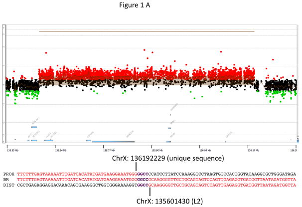
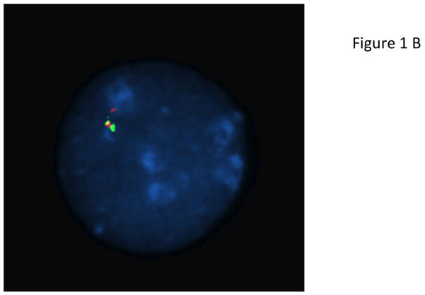
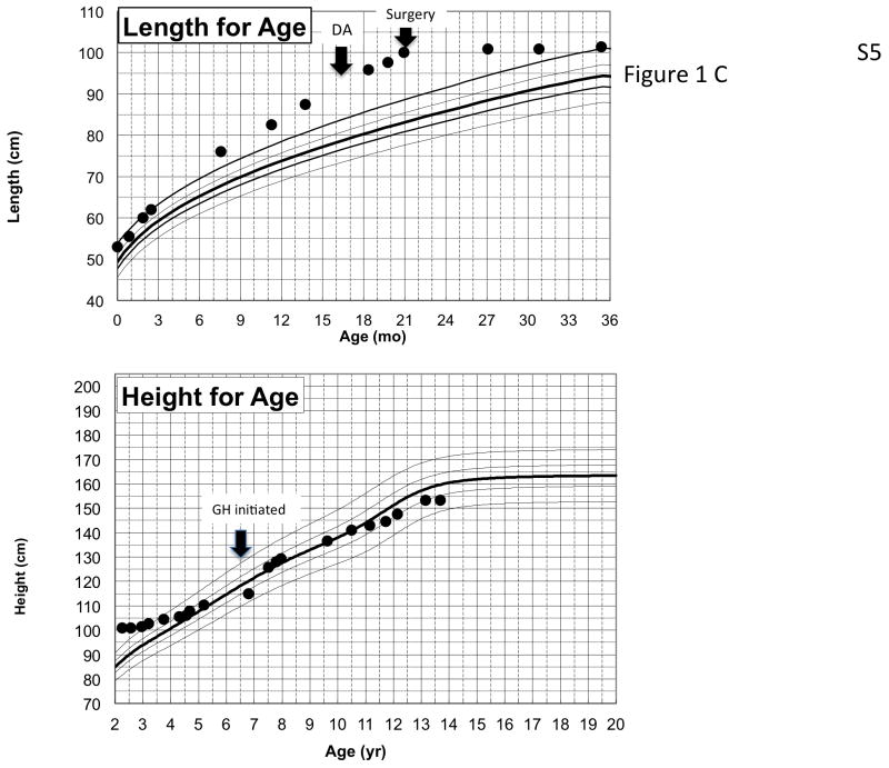
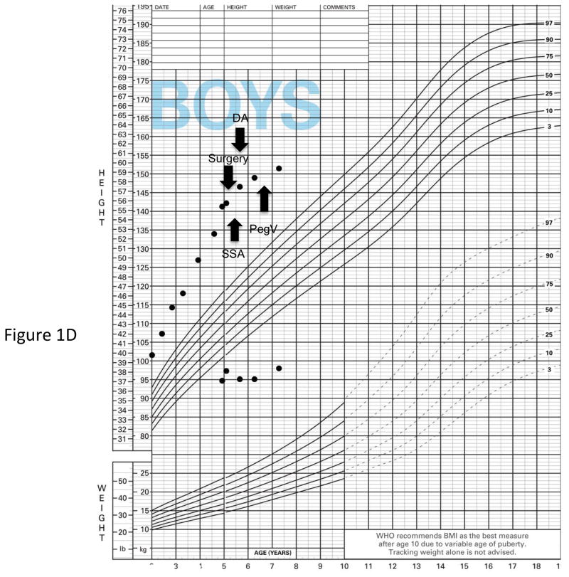
Figure 1.
Genetic and growth features in patients with X-LAG syndrome. Panel A Representative chromosome Xq26.3 microduplication in female sporadic patient S10 on HD-CGH array showing the duplicated region (red) and details of the breakpoint regions of microhomology leading to the duplication (below in purple GGCC). FISH results for sporadic patient S1 (male) showing a duplication of the proximal and distal probes (red/green) in a representative leukocyte. Growth patterns during early life in sporadic patients S5 (female) and S1 (male). Panel C shows the growth in patient S5 that exceeded the 97 centile for length as early as 6 months of age; she was treated with cabergoline DA and then surgery, after which time the total resection of tumor was associated with flattening of her growth curve. She eventually was diagnosed with panhypopituitarism and diabetes insipidus and required GH supplementation as indicated. Panel D shows the early growth of patient S1, a sporadic male case that began between the ages of 9 and 12 months and continued despite subtotal surgical resection, SSA and DA; the introduction of pegvisomant (PegV) at the age of 6 led to a decrease in gain in height.


