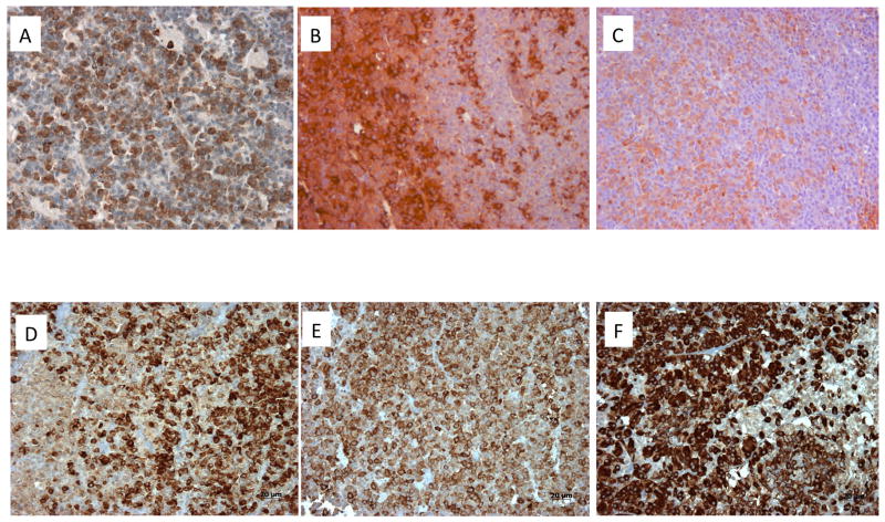
Figure 5.
Immunohistochemistry of pituitary adenomas from Xq26.3 microduplication cases. In case S1 (sporadic male) Panel A demonstrates strong GHRH-R staining (brown) in the pituitary adenoma using a C-terminal GHRH-R peptide (20× magnification). The tumor was a mixed GH and PRL adenoma and staining of consecutive slices demonstrated largely different cell populations that stained for GH (brown; Panel B) and PRL (brown; Panel C). SSTR immunohistochemistry was also performed on tumors from patients with the Xq26.3 microduplication. AIP staining (brown) shown in 3 pituitary tumors from X-LAG syndrome cases (Panels D–F)). In all 6 cases tested AIP staining was preserved at moderate to high levels and was predominantly cytoplasmic (20× magnification).

