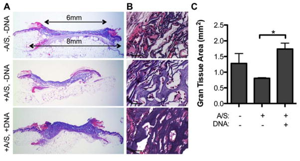
Figure 1.

H&E stained full wound cross-sections (A, 1.6x magnification) with corresponding 40x magnification images (B, scale bar = 50 μm) of wounds with 100 μm porous hydrogels either with or without agarose/sucrose and/or pDNA/PEI polyplexes. (C) Quantification of granulation tissue (n = 3 – 4) reveals differences between granulation tissue formation surrounding porous hydrogels with various matrix components after 14 days of healing. A/S = agarose/sucrose.
