Figure 4.
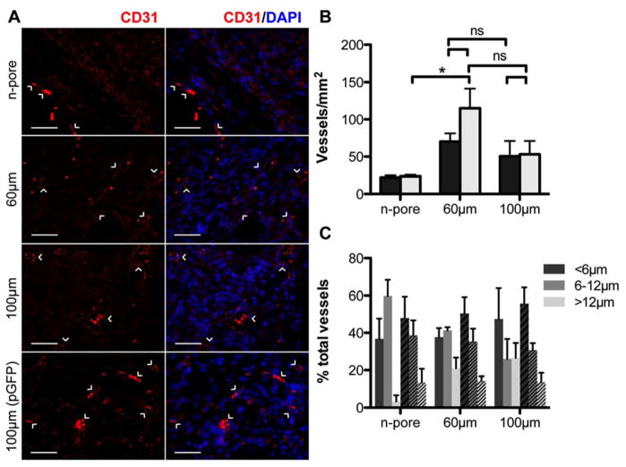
Staining for endothelial marker CD31 confirms the presence of small capillaries within the granulation tissue of 60 and 100 μm porous hydrogel implants loaded with pVEGF (A, scale bar = 50 μm; PECAM positive staining (CD31+ endothelial cells) = red, cell nuclei = blue). 100 μm porous hydrogel implants loaded with pGFPluc are shown for comparison. Quantification of vessel density (n = 4 – 5) reveals a slight correlation to hydrogel porosity (B, black bars = pVEGF, grey bars = pGFPluc loaded hydrogels). Vessel diameters were also measured (C, solid bars = pVEGF, striped bars = pGFPluc loaded hydrogels). Approximately 40–50 % of vessels in all samples were less than 6 μm in diameter.

