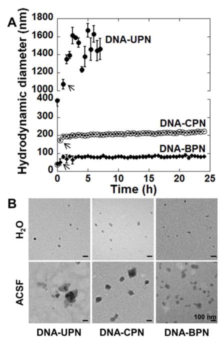
Figure 1. Stability of gene vector in artificial cerebrospinal fluid.
(A) Hydrodynamic diameter of gene vectors in aCSF at 37 °C was measured by dynamic light scattering (DLS). Measurements were taken every 30 min up to 24 hours until polydispersity (PDI) > 0.5. Data represents the mean ± SEM. Note that hydrodynamic diameters of DNA-CPN and DNA-BPN are overlapping at 0 h. Arrows denote hydrodynamic diameters of DNA-UPN, DNA-CPN and DNA-BPN at 30 min post-incubation. (B) Transmission electron microscopy images of gene vectors in ultrapure water (top panel) and following 1 h incubation in aCSF at 37 °C (bottom panel). Scale bars = 100 nm.

