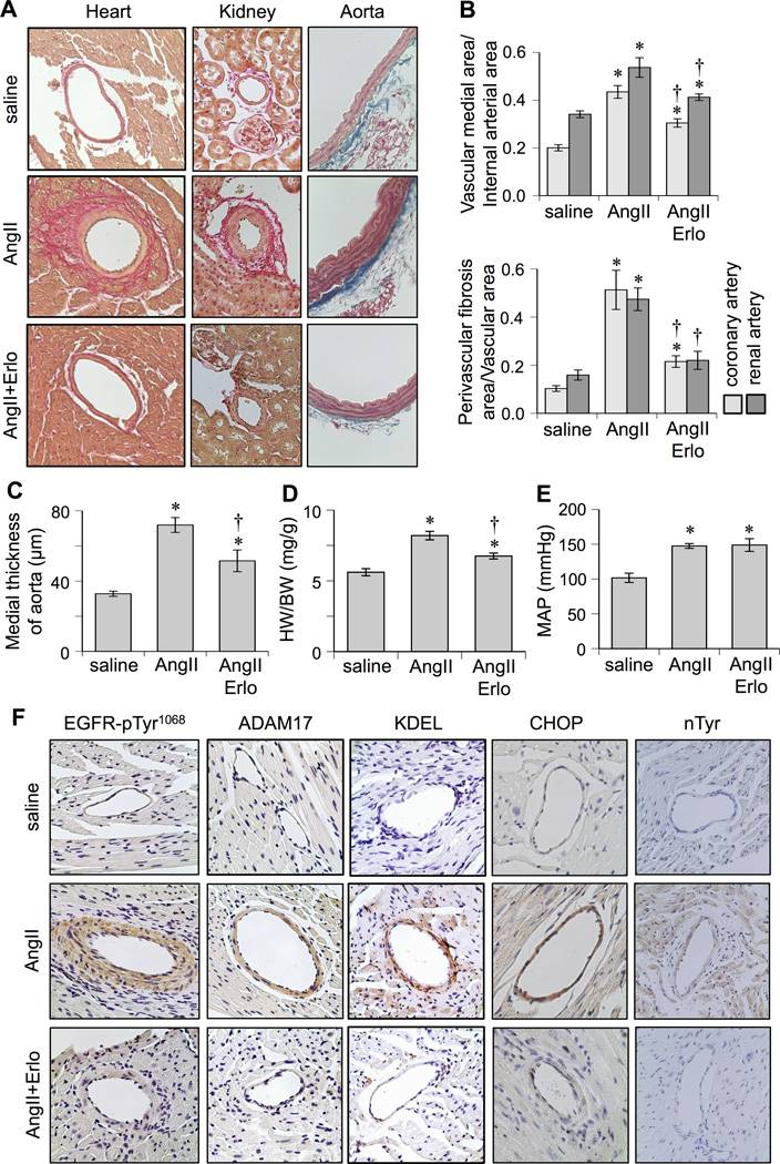
Figure 1.
Effects of EGFR inhibitor, erlotinib, on cardiovascular remodeling induced by AngII. C57Bl/6 mice were infused with saline (n=8) for 2 weeks, or AngII (1 µg/kg/min) for 2 weeks with (n=8) or without (n=8) treatment of erlotinib (10 mg/kg/day intraperitoneal injection). Hearts and kidneys were stained with Sirius red and aortas were stained with Masson trichrome (Mean±SEM). A: Representative staining (200x) is presented. B: Quantification of medial area to internal arterial area of the coronary and renal arteries, and quantification of perivascular fibrosis area to vascular area of these arteries. C: Quantification of medial thickness of the thoracic aorta. D: Heart weight (HW) body weight (BW) ratio. E: Mean arterial pressure (MAP) was evaluated by telemetry. F: Heart sections were immuno-stained with antibodies as indicated (n=4). Antibodies against KDEL and CHOP were used to assess ER stress. Antibody against nitro-tyrosine (nTyr) was used to assess oxidative stress. *p<0.05 compared with control saline infusion. †p<0.05 compared with AngII infusion.

