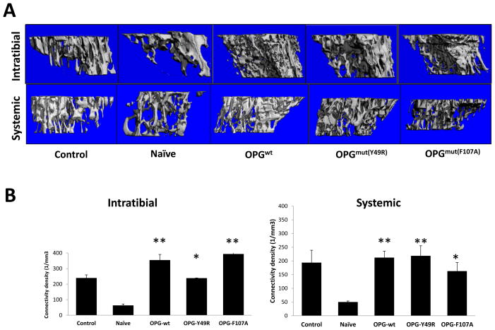
Figure 5. Micro-CT assessment to determine the potential of OPGmut in inhibiting tumor-induced osteolytic damage.
Nude mice were injected intratibially with ~1×10⁵ osteolytic prostate cancer cell line PC3. Twenty-four hours later, ~3×10⁵ MSC over-expressing OPGwt, OPGmY49R or OPGmF107A were injected into the same tibia for intratumoral administration and by tail vein injection for systemic administration. Mice from both routes of MSC administration were sacrificed 14 days post MSC therapy and tibia were collected for Micro-CT analysis to determine changes in the overall trabecular architecture (A) and connectivity density (B) (*p<0.05; **p<0.01, compared to Naïve).

