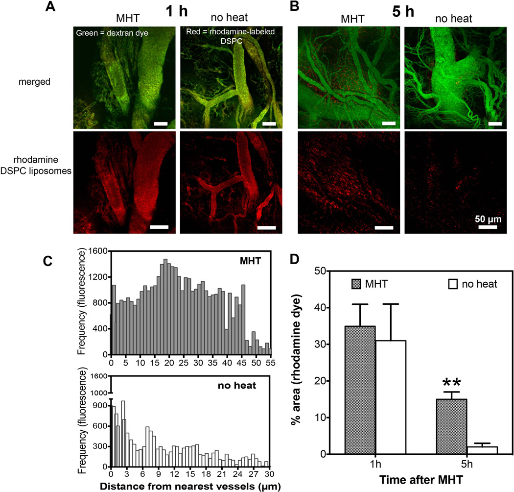
Figure 4. MHT treatment enhances accumulation and spatial distribution of nanoliposomes.
A) Intravital microscopy images obtained 1 h after MHT treatment showing nanoliposomes (red); the liposomal accumulation in treatment group was indistinguishable from control group where nanoparticles remained in circulation (green, vessels; B) while analyses at 5 h after treatment revealed significant nanoparticle accumulation (red) in tumor interstitial space compared to untreated group; C) further analyses showed increased nanoparticle spatial distribution with tumor receiving MHT treatment showing liposomal dispersion of 21 ± 2.5 µm away from the nearest tumor vessels versus 3.7 ± 1.9 µm for untreated tumors; D) MHT treatment showed 3-fold increase in accumulation of nanoparticles at 5h and no significant enhancement at 1 h after treatment.

