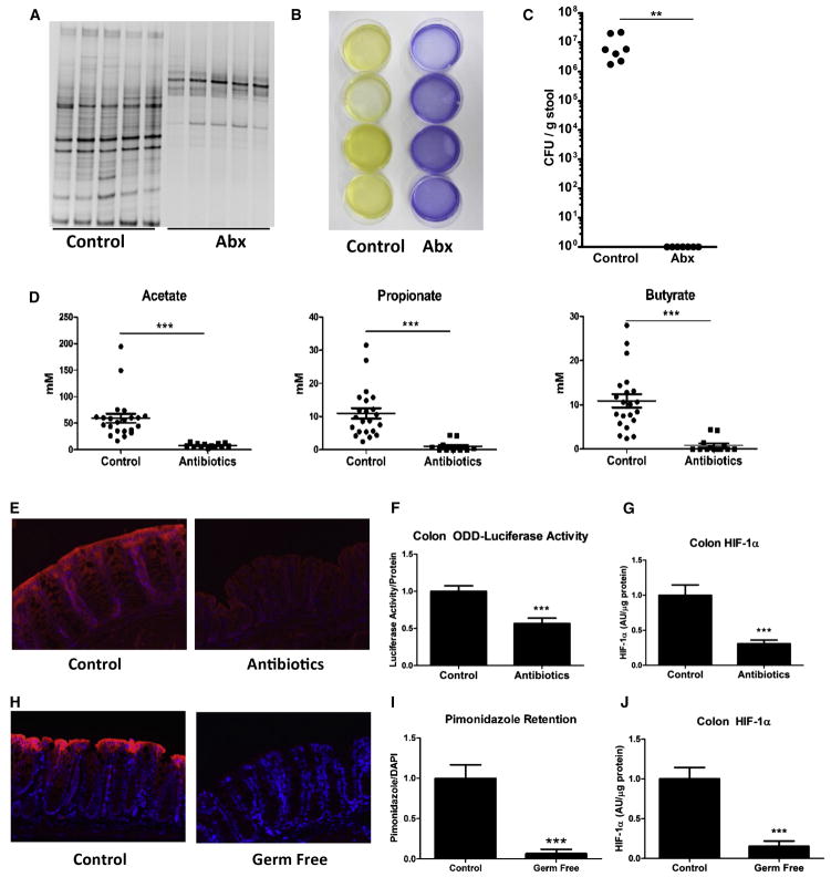
Figure 3. Antibiotics Reduce SCFA Concentration and HIF in the Colon.
(A) Denaturing gradient gel electrophoresis, where each lane represents an individual animal and each band represents a distinct bacterial population, revealed depletion of most bacterial populations in the cecum of ODD-Luc mice with Abx treatment.
(A and B) Despite appearance of resistant organisms (A), cecal contents from Abx-treated mice lost their ability to ferment inulin to SCFA. Each plate contained cecal contents from an individual animal, and yellow color indicates pH < 5.2 (B).
(C) Analysis of culturable stool microbiota from vehicle and Abx-treated mice (n = 7 per group, where ** indicates p < 0.025).
(D) Accordingly, cecal acetate, propionate, and NaB were reduced with Abx treatment (p < 0.001).
(A–C) Data represent average ± SEM from three independent experiments ([A] and [B]). Each lane (A), dish (B), and symbol (C) represent an individual animal.
(E) Significance was determined using Student’s t test. Abx treatment reduced retention of PMDZ in the colon indicating loss of “physiologic hypoxia.”
(F and G) This influence reflected reduced luciferase activity ([F], p < 0.001) and HIF-1α protein ([G], p < 0.001) in the colon of ODD-Luc animals.
(H) PMDZ localization in control and germ-free mouse colon (n = 6 per group). PMDZ was quantified (H) where *** indicates p < 0.001.
(J) HIF-1α protein in control and germ-free mouse colon (n = 6 per group). Data represent average ± SEM from three independent experiments where *** indicates p < 0.001.

