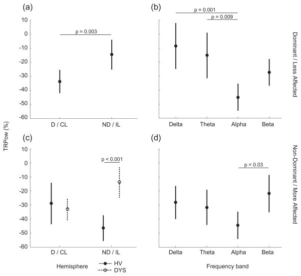
Figure 4.
Mean task-related EEG spectral power change (TRPow) in each hemisphere (a and c) and in each frequency band (b and d). Data from the dominant/less affected wrist are in the top row (a and b); data from the non-dominant/more affected wrist are in the bottom row (c and d). P values for all statistical differences between factors are indicated. Bars represent the 95% confidence interval. Closed circles represent HV and open circles represent DYS in order to show the group-hemisphere interaction on TRPow from the Non-dominant/more affected wrist task. D/CL = Dominant/Contralesional hemisphere, ND/IL = Non-dominant/Ipsilesional hemisphere, HV = Healthy volunteers, DYS = Dystonia group.

