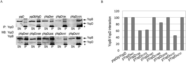
Fig 5. Interaction of the different YopD mutants with YopB.

A. YopB-YopD complexes secreted in bacterial supernatants of yopD, yopD/pYopD and yopD expressing the different yopD mutants were precipitated by 1 h incubation with YopD mab (clone 248:19) followed by incubation with Dynabeads Protein G (Invitrogen). After 3 washes, beads were resuspended in 2X Laemmli sample buffer, and boiled. The eluted material (IP) and an aliquot of the bacterial supernatants (SN) were resolved in SDS-PAGE. Western blot was performed using anti-YopD and anti-YopB Mabs, and anti-mouse IR680 or IR800 secondary antibodies. Bands corresponding to YopB and YopD are indicated by arrows. Also present in the IP samples are a band originated from the beads (marked with an asterix), and a weak band of unknown origin that migrates right below YopB. B. Signal intensities of immunoprecipitated YopB and YopD were calculated using Odyssey imaging system software (LI-COR Biosience). YopB-YopD interaction for each mutant was calculated as the ratio between immunoprecipitated YopB and YopD. Results were normalized to yopD/pYopD.
