Figure 1.

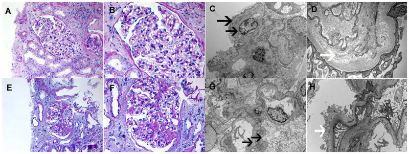
Kidney biopsies. Biopsy 1 (Top panel): A–B. Light microscopy showing segmental thickening of the glomerular basement membranes (Perioidic acid Schiff stain; original magnification, 20× and 40×, respectively). Electron microscopy showing (C) double contour formation along capillary walls (4200x), and (D) subendothelial expansion with fluffy granular material (33000x). Biopsy 2 (bottom panel): E–F Light microscopy showing segmental sclerosis (Perioidic acid Schiff stain; original magnification, 20× and 40×, respectively). Electron microscopy showing (G) double contour formation along capillary walls (5000x), and (H) subendothelial expansion with fluffy granular material (12000x). Black arrows, double contours; white arrows, subendothelial fluffy material.
