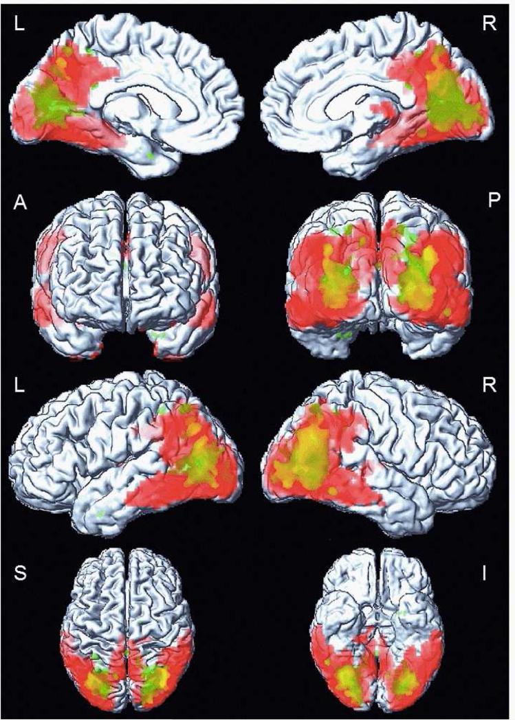
Figure 4. Overlapping patterns of white matter and cortical grey matter alterations in DLB patients on voxel-based analysis.
Cortical glucose hypometabolism on 18F-FDG PET is overlapping with reduced FA in white matter on DTI in DLB patients compared to CN. The overlap is displayed on a surface render using SPM5. The white matter alteration (green color) is confined to parieto-occipital region, p<0.001 (uncorrected). The alteration in cortical glucose metabolism (red color) is more diffuse, in posterior temporal, parietal and in occipital cortices, p<0.001, (FWE corrected).

