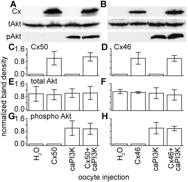
Figure 4. Injection of oocytes with caPI3K increased activation of Akt without effecting connexin or t-Akt protein levels.
(A) Immunoblotting confirmed the expression of Cx50 and increased levels of p-Akt, with no change in t-Akt levels between all of the samples. (B) Expression of Cx46 did not change when co-expressed with caPI3K. (C) Densitometry measurements (averages of three independent experiments) showed there was no significant difference in Cx50 protein when co-expressed with caPI3K. (D) Quantification of Cx46 protein levels confirmed they were not affected by Akt activation. (E), (F) Total Akt expression is used as a control. (G) (H) Quantification of p-Akt bands verified equal activation of Akt in oocytes co-expressing caPI3K and connexin proteins.

