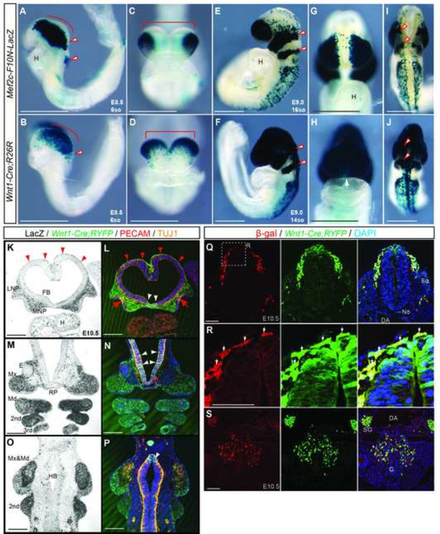
Fig.5. Mef2c-F10N-LacZ marks cells in a pattern very similar to Wnt1-Cre but is specific for migratory NCC.
(A–J) Comparison between Mef2c-F10N-LacZ (A, C, E, G, I) expression and Wnt1-Cre;R26R lineage marking (B, D, F, H, J) in whole embryos. Distribution of β-galactosidase activity in lateral (A, B, E, F), frontal (C, D, G, H) and dorsal (I, J) views of E8.5, 6-somite stage (A–D) and E9.0, 16-somite stage (E–J) embryos. Red outline arrowheads indicate pre-otic NCC streams. (K–P) Frontal sections of E10.5 Mef2c-F10N-LacZ and Wnt1-Cre;Rosa-YFP compound embryos. Mef2c-F10N-LacZ expression is revealed by β-galactosidase activity (K, M, O), and Wnt1-Cre;YFP lineage marking is revealed by immunostaining for YFP in conjunction with the endothelial marker CD31, and the neural marker Tuj1. Red arrowheads indicate NCC of dorsal forebrain that may contribute to the meninges. Red tailed arrows indicate delaminating olfactory nerve from the olfactory epithelium. White arrows indicate the overlap with migrating NCC. (Q–S) Double staining of β-galactosidase expression from the Mef2c-F10N-LacZ transgene and YFP expression from Wnt1-Cre;Rosa26YFPi+ in neural tube (Q, R) and gut (S) in E10.5 embryos. (R) High magnification images of white dashed inset in (Q). White arrows indicate the overlap with migrating NCC. DAPI is used to stain nuclei. Abbreviations: 2nd second pharyngeal arch; 3rd pharyngeal arch; DA, dorsal aorta; E, eye; FB, forebrain ventricle; G, gut; H, heart; HB, hindbrain; LNP, lateral nasal process; Md, mandibular process; Mx, maxillary process; MNP, medial nasal process No, notochord; RP, Ratke’s pouch; So, somite; SG, sympathetic ganglion; 3rd, third pharyngeal arch; TG, trigeminal ganglion. Scale bars: 0.5 mm in A–P, 0.1 mm in Q–S.

