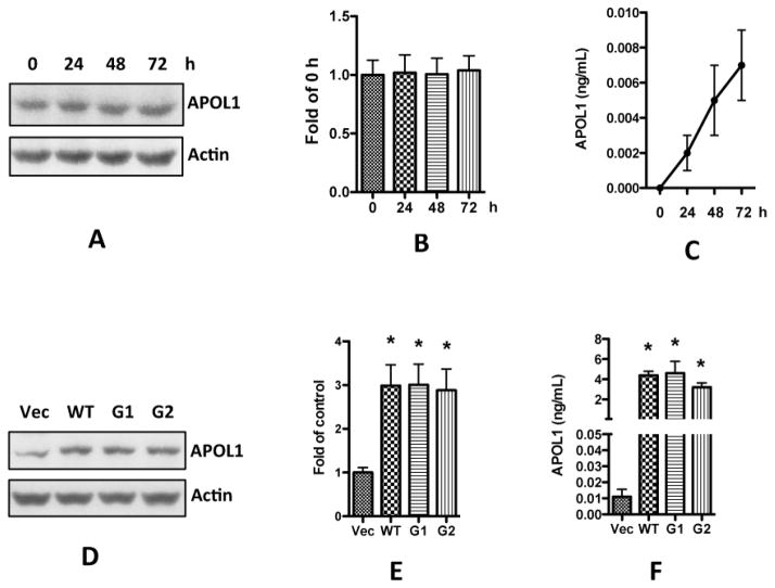
Figure 5. Overexpression of APOL1 in human vascular smooth muscle cells.
A–C: HSMCs were cultured in SMCM until reaching 80–90% confluence, and were transferred to RPMI medium. After another 24, 48, and 72 h, cell lysates and medium were collected for detection of APOL1 with Western blots (A-B) and ELISA (C), respectively.
D–F: HSMCs were infected with lentivirus (titrated as 0.4 pg HIV p24 protein per cell) for 3 h, and then re-incubated in fresh medium for 48 h (n=3). Cells and media were collected.
D. Protein blots were probed for APOL1, stripped and re-probed for actin.
E. Densitometric analysis shown as bar graphs. * p < 0.05 compared with control (vector).
F. APOL1 concentrations were determined in media with an ELISA kit.
Results (mean ± SD) are from three sets of experiments carried out in triplicate. * p < 0.05 compared with control (vector). Vec, vector alone; WT, APOL1 wild type; G1, APOL1 G1 variant; G2, APOL1 G2 variant.

