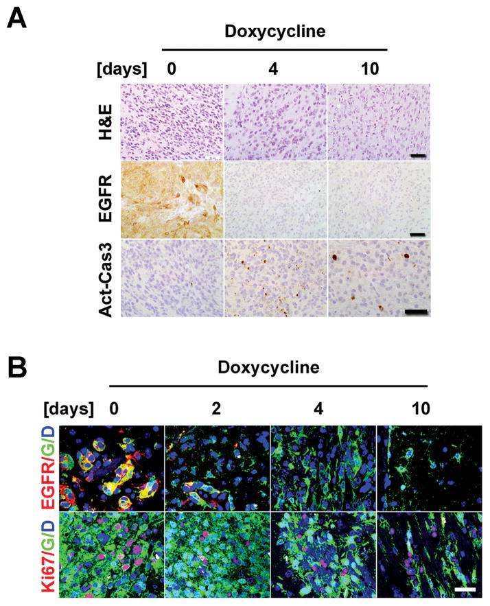
Figure 5.
EGFR*-independent glioma cells exist prior to treatment. A, tumor bearing animals grafted with GFP-expressing iEIP glioma cells were switched to Dox and sacrificed at indicated time-points (n = 2 for each). H&E and IHC staining against EGFR or activated caspase3 (Act-Cas3) revealed complete suppression of EGFR* induction at 4-day post treatment and enhanced apoptosis in Dox treated tumors. Scale bars represent 50 μm. B, IF staining revealed a subpopulation of GFP-labeled Ki67-positive proliferative tumor cells persisted through genetic suppression of EGFR* induction. G - GFP; D - DAPI. Scale bars represent 100 μm.

